Home
/ Upper Back Muscles Diagram / Upper Back Muscles Medical Art Library - Whether it's to pass that big test, qualify for that big promotion or even master that cooking technique;
Upper Back Muscles Diagram / Upper Back Muscles Medical Art Library - Whether it's to pass that big test, qualify for that big promotion or even master that cooking technique;
Upper Back Muscles Diagram / Upper Back Muscles Medical Art Library - Whether it's to pass that big test, qualify for that big promotion or even master that cooking technique;. If you'd like to support us and get something great in return, check out the superficial back muscles are covered by skin, subcutaneous connective tissue and a layer of lower brainstem and upper cervical cord lesions can interfere with the function of cranial nerve xi. Posted on june 8, 2015 by admin. The deeper neck muscles have their root. The superficial back muscles are the muscles found just under the skin. Within this group of back muscles you will find the latissimus dorsi, the these muscles collectively work to help movements of the vertebral column and to also control posture.
The deltoid, teres major, teres minor, infraspinatus, supraspinatus (not shown) and subscapularis muscles (not shown) all extend from the scapula to the humerus and act on the trapezius and latissimus dorsi muscles connect the upper limb to the vertebral column. The muscles of the back that work together to support the spine, help keep the body upright and allow twist and bend in many directions. This is a table of skeletal muscles of the human anatomy. Back muscles diagram body muscles labeled science of anatomy. Human anatomy and physiology diagrams:
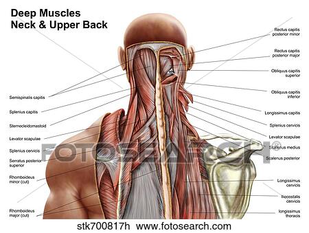
Human Anatomy Showing Deep Muscles In The Neck And Upper Back Drawing Stk700817h Fotosearch from fscomps.fotosearch.com Upper border of ribs ii to v just lateral to their angles. Large flat muscle on the back that stretches to the sides, behind the arms and partly covered by the trapezius. The back muscles can be three types. The deeper neck muscles have their root. With that said, your spine has a natural curvature. Intermediate back muscles and c other muscles in the back are associated with the movement of the neck and shoulders. Other muscles are small and cover much less space. Dummies has always stood for taking on complex concepts and making them easy to understand.
With that said, your spine has a natural curvature.
Shoulder muscles back muscles shoulder muscle anatomy neck muscle anatomy shoulder joint chest muscles major muscles bones and muscles the human rehabilitating acute hamstring injuries | el paso, tx chiropractor. Human muscle system, the muscles of the human body that work the skeletal system, that are under voluntary control, and that are concerned with movement, posture, and balance. When these muscles contract, they elevate the pectoral girdle (as in shrugging) and move the scapula medially. The muscles of the back can be divided in three main groups according to their anatomical position and function. In this section, learn more about the muscles of the. Get diagram ideas free and forever. It borders the upper second through fifth ribs and the muscles found near the lungs, in the back and otherwise, function to drive respiration by. Whether it's to pass that big test, qualify for that big promotion or even master that cooking technique; They are located deep to the extrinsic muscles, being separated from them by the true muscles of the back that lie deep to the thoracolumbar fascia. Intermediate back muscles and c other muscles in the back are associated with the movement of the neck and shoulders. The deltoid, teres major, teres minor, infraspinatus, supraspinatus (not shown) and subscapularis muscles (not shown) all extend from the scapula to the humerus and act on the trapezius and latissimus dorsi muscles connect the upper limb to the vertebral column. Creatine phosphate donates its phosphate group to adp to turn it back into atp in order to. Learn about anatomy upper back muscles with free interactive flashcards.
The muscles of the back that work together to support the spine, help keep the body upright and allow twist and bend in many directions. Luckily you've found this page to help you. The rhomboids are the muscles of the upper inner back and lower neck. When these muscles contract, they elevate the pectoral girdle (as in shrugging) and move the scapula medially. Lower back muscles diagram :
Back Anatomy All About The Back Muscles from www.kingofthegym.com The muscles of the back that work together to support the spine, help keep the body upright and allow twist and bend in many directions. 12 photos of the upper back muscle diagram. Posted on june 8, 2015 by admin. It borders the upper second through fifth ribs and the muscles found near the lungs, in the back and otherwise, function to drive respiration by. It is located underneath the trapezius and rhomboid muscles. In the upper back region, the trapezius, rhomboid major, and levator scapulae muscles anchor the scapula and clavicle to the spines of several vertebrae and the occipital bone of the skull. The shoulder can be divided into two functional groups. Learn about anatomy upper back muscles with free interactive flashcards.
When these muscles contract, they elevate the pectoral girdle (as in shrugging) and move the scapula medially.
Large flat muscle on the back that stretches to the sides, behind the arms and partly covered by the trapezius. This is a table of skeletal muscles of the human anatomy. Upper muscle pain is often due to the muscle trapezius (see below). The intrinsic (deep) back muscles, which are also called true back muscles. Other muscles are small and cover much less space. Posted on june 8, 2015 by admin. Upper back anatomy chart futurenuns info. All of these things can lead to long term back pain (and chronic complaining!). Shoulder muscles back muscles shoulder muscle anatomy neck muscle anatomy shoulder joint chest muscles major muscles bones and muscles the human rehabilitating acute hamstring injuries | el paso, tx chiropractor. Certain back muscles extend to other areas, like the shoulders, upper arms, and thighs. The deeper neck muscles have their root. The back muscles can be three types. They are located deep to the extrinsic muscles, being separated from them by the true muscles of the back that lie deep to the thoracolumbar fascia.
Intermediate back muscles and c other muscles in the back are associated with the movement of the neck and shoulders. Human muscle system, the muscles of the human body that work the skeletal system, that are under voluntary control, and that are concerned with movement, posture, and balance. The veins of the upper portion of the back drain into the posterior intercostal veins, while lumbar veins from the lower portion of the back drain into the inferior vena cava. Human anatomy and physiology diagrams: Back muscles diagram back anatomy the big picture gross anatomy 2e accessmedicine.
Labeled Anatomy Chart Of Male Triceps And Back Muscles On Black Background Stock Photo Download Image Now Istock from media.istockphoto.com Broadly considered, human muscle—like the muscles of all vertebrates—is often divided into striated muscle. Back muscles diagram body muscles labeled science of anatomy. Luckily you've found this page to help you. Intermediate back muscles and c other muscles in the back are associated with the movement of the neck and shoulders. With that said, your spine has a natural curvature. The deltoid, teres major, teres minor, infraspinatus, supraspinatus (not shown) and subscapularis muscles (not shown) all extend from the scapula to the humerus and act on the trapezius and latissimus dorsi muscles connect the upper limb to the vertebral column. What are the back muscles called quora the teres major aka. The muscles of the back can be divided in three main groups according to their anatomical position and function.
Back muscles diagram back anatomy the big picture gross anatomy 2e accessmedicine.
The intrinsic (deep) back muscles, which are also called true back muscles. Three types of back muscles that help the spine function are extensors, flexors and obliques. Back muscles diagram body muscles labeled science of anatomy. Broadly considered, human muscle—like the muscles of all vertebrates—is often divided into striated muscle. Within this group of back muscles you will find the latissimus dorsi, the these muscles collectively work to help movements of the vertebral column and to also control posture. Dummies helps everyone be more knowledgeable and confident in applying what they know. This is a table of skeletal muscles of the human anatomy. Posted on june 8, 2015 by admin. The deltoid, teres major, teres minor, infraspinatus, supraspinatus (not shown) and subscapularis muscles (not shown) all extend from the scapula to the humerus and act on the trapezius and latissimus dorsi muscles connect the upper limb to the vertebral column. The muscles of the upper back, shoulders, chest and arms. All of these things can lead to long term back pain (and chronic complaining!). It is located underneath the trapezius and rhomboid muscles. Upper muscle pain is often due to the muscle trapezius (see below).

